ShanghaiDoctor - Where China's Healing Wisdom Shapes Modern Medicine

Update time:2026-02-02Visits:3621
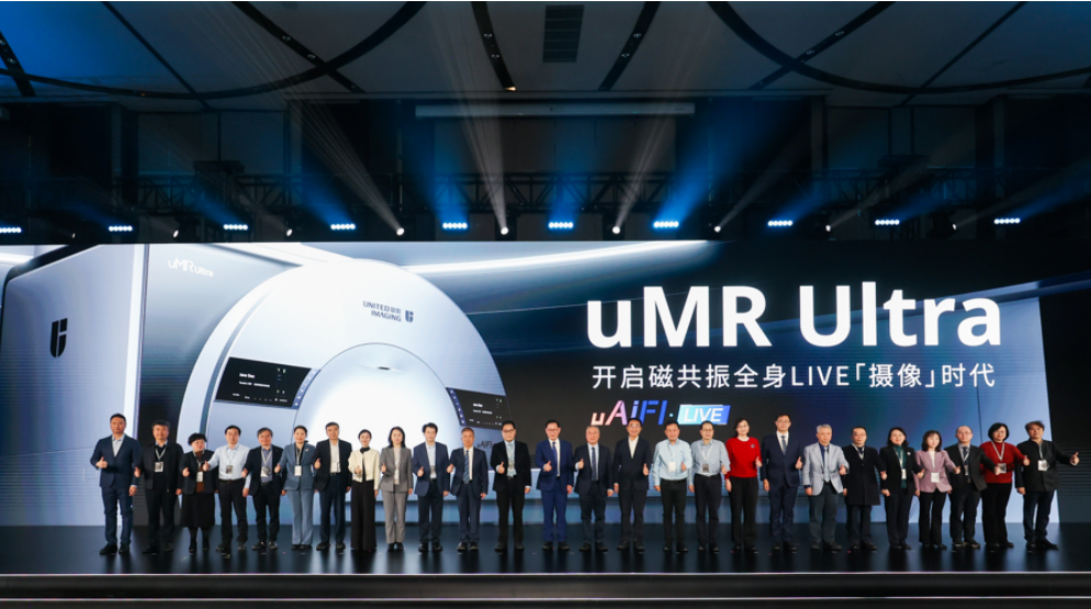
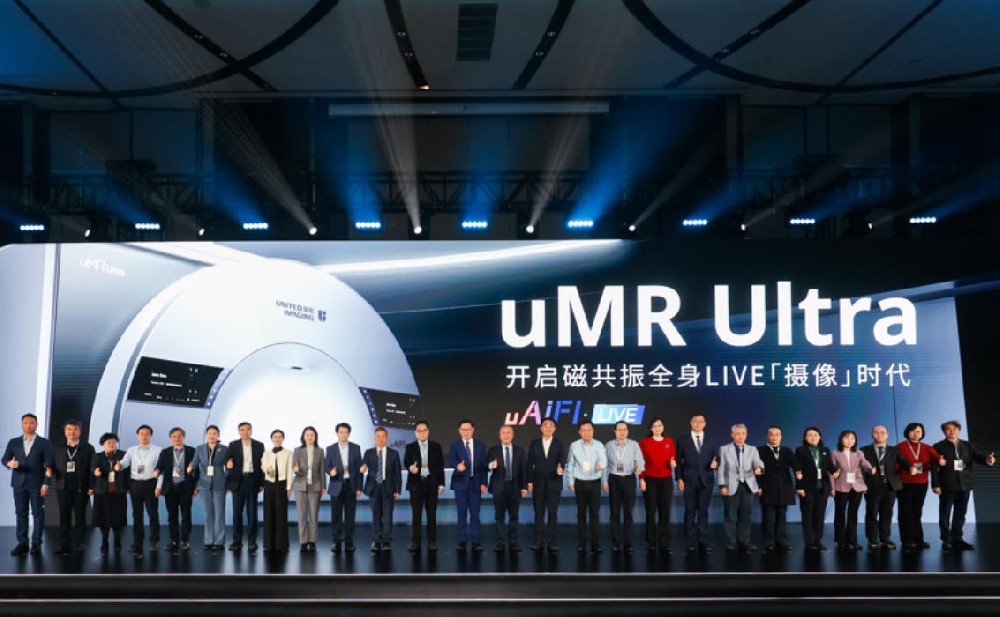
SHANGHAI, Feb. 1, 2026 — Reported by ShanghaiDoctor.cn — The 2026 United Imaging MRI Innovation Conference, jointly hosted by the National Innovation Center for High-Performance Medical Devices and United Imaging Healthcare (UIH), commenced today in Shanghai. At the event, UIH officially launched the world’s first high-definition “video camera” MRI system. This revolutionary technology is hailed as a breakthrough that transitions magnetic resonance imaging from an era of static “photography” to dynamic “cinematography,” holding immense potential for the observation, diagnosis, and research of moving human body parts.
Top-tier Chinese medical institutions, including Peking Union Medical College Hospital (PUMCH), Huashan Hospital affiliated with Fudan University, and Renji Hospital affiliated with Shanghai Jiao Tong University School of Medicine, are among the first to adopt the technology. This marks a critical milestone where original Chinese imaging technology is entering a key phase of large-scale clinical validation.
AI-Empowered Physiological Imaging: Chinese Innovation Leads the Globe
MRI has long been a cornerstone of clinical diagnosis. However, traditional MRI acquisition produces images similar to a series of static photographs, primarily reflecting the morphological structure of tissues and organs within a specific timeframe. This approach often faces limitations when observing life activities such as heartbeats, breathing, gastrointestinal peristalsis, and cerebrospinal fluid (CSF) flow.
The introduction of UIH’s LIVE technology represents a fundamental transformation for the industry. As the industry’s first high-definition dynamic imaging technology, it marks a paradigm shift in medical imaging from "seeing structure" to "understanding process."
Professor Liang Dong from the Shenzhen Institutes of Advanced Technology (SIAT) at the Chinese Academy of Sciences explained that traditional rapid imaging indirectly presents "motion" by combining multiple static frames after acquisition, which often fails when facing irregular complex movements like gastrointestinal peristalsis. The core of LIVE dynamic imaging lies in utilizing prior information, such as the inherent movement patterns of organs, to actively and precisely characterize dynamic processes. It breaks through traditional algorithm frameworks by innovating at the foundational level, thereby capturing irregular complex physiological movements.
“Artificial intelligence is propelling MRI into a new stage of intelligent physiological imaging,” said Professor Yan Fuhua, Director of the Department of Radiology at Ruijin Hospital and Head of the MRI Group of the Chinese Society of Radiology. “In the past, we always tried to make the human body ‘static’ to obtain clear images. Now, dynamic MRI can truly restore natural physiological activities, allowing doctors to capture disease clues in motion. This not only opens new avenues for research into CSF circulation and gastrointestinal motility but also provides a vital platform for establishing new diagnostic standards and research systems.”
On September 29, 2025, the uMR Ultra, the world’s first “video camera” MRI independently developed by UIH, officially received market approval from the National Medical Products Administration (NMPA).
“For the past hundred years, we have been solving the problem of ‘seeing and seeing clearly.’ But the essence of life is eternal rhythm,” said Academician Wang Zhenchang in his conference address. “CSF circulation, heart beating, gastrointestinal functioning—these dynamic processes were precisely the blind spots in past diagnoses. Today, this technology allows us not to just look at a photo, but to possess a complete segment of ‘film.’ This is a qualitative leap.”
Validation by Top Hospitals: Dynamic Imaging Reshapes Diagnosis
Currently, the uMR Ultra has been systematically deployed at Peking Union Medical College Hospital, Huashan Hospital, and Renji Hospital. In scenarios such as abdominal and pelvic functional assessment, CSF dynamics research, and the diagnosis of gastrointestinal and biliopancreatic diseases, LIVE imaging has significantly improved lesion detection rates.
At Huashan Hospital, the system is being used systematically for nervous system disease research. By establishing new quantitative analysis paths centered on CSF dynamics, it provides more precise evidence for pre- and post-operative evaluation of conditions like spinal canal stenosis and Chiari malformations.
“Dynamic imaging allows us to observe phenomena that were previously uncatchable, providing significant guidance for disease diagnosis, treatment decisions, and efficacy assessment,” said Yao Zhenwei, Director of the Department of Radiology at Huashan Hospital.
In the past, for patients with CSF leaks, doctors often knew the disease existed but not its location. Clinically, leaks could be inferred from symptoms like intracranial hypotension, but finding the tiny “leak point” was like finding a needle in a haystack. Diagnosis required a series of complex checks with limited detection rates. Today, using this new MRI, hospital teams can clearly see the dynamic process of cerebrospinal fluid flowing from the leak point on the screen, quickly locking onto the lesion.
“We used to be followers, and suddenly we are ‘leaders,’” Yao Zhenwei stated frankly. “The significant meaning of this technology is that original Chinese technology is truly moving toward international leadership. Facing this rare opportunity, the research team has a strong sense of mission, striving to thoroughly master the technology and solidify the results as quickly as possible.”
At Renji Hospital, the equipment has been introduced for the diagnosis of gastrointestinal, biliopancreatic, and urinary system diseases, particularly in complex scenarios such as inflammatory bowel disease and periampullary tumors.
Zhou Yan, Director of the Department of Radiology at Renji Hospital, noted that in clinical practice, the changes brought by “video camera” MRI technology are obvious at a glance. In intestinal examinations, it not only clearly distinguishes between lesions and normal peristalsis but also significantly shortens scanning time.
Zhou Yan added that beyond evaluating Crohn’s disease and intestinal obstructions in the digestive system, LIVE technology has shown broad application prospects. The team has expanded its use to autoimmune diseases, such as intestinal issues caused by systemic sclerosis, the evaluation of outlet constipation, and even the prostate field. Excellent application value has also been discovered in the diagnosis of bladder cancer and ureteral cancer in the urinary system.
At Peking Union Medical College Hospital (PUMCH), the uMR Ultra focuses on high-motion areas such as the abdomen and pelvis. Through continuous imaging, doctors can simultaneously observe structural details and functional activities, allowing for a more comprehensive assessment of pelvic floor disorders, intestinal diseases, and complex post-operative states.
The World’s First Invasive Brain-Computer Interface Just Got Approved in Shanghai
A Shanghai Hospital Launches Vertical Large Model for Women’s Health
Shanghai Research Team Develops AI Diagnostic System for Rare Diseases with “Explainable” Reasoning
“Yi Jian Kang” Foundation Established by Professor Wen Yumei Celebrates 13 Years of Impact
Shanghai Hosts Landmark Global Transplant Congress, a First for Mainland China
The World’s First Invasive Brain-Computer Interface Just Got Approved in Shanghai

A Shanghai Hospital Launches Vertical Large Model for Women’s Health
Shanghai Research Team Develops AI Diagnostic System for Rare Diseases with “Explainable” Reasoning
World’s First “Video Camera” MRI is set in Shanghai, Ushering in the Era of Dynamic Physiological Imaging

“Yi Jian Kang” Foundation Established by Professor Wen Yumei Celebrates 13 Years of Impact

Renji International Medical Marks 20th Anniversary

Shanghai Overseas Patients See 25% Growth

Shanghai Hosts Landmark Global Transplant Congress, a First for Mainland China

Shanghai Health Report, Issue 7: Expert Guidance

Shanghai Doctors Become Viral Health Educators
Copyright © 2023 Yewen Renyi & ShanghaiDoctor.cn (This website is a non - profit medical humanities platform. The information contained herein is solely for biographical and historical purposes and does not constitute any medical advice.) 沪ICP备2023005392号-2 XML 沪公网安备31010902100835号
